

MADRID, MADRID, SPAIN, March 14, 2024 /EINPresswire.com/ -- Scientists are determined to tackle the complexities of Alzheimer’s disease, a condition known for its devastating impact on memory and cognitive function. A new study presents an innovative method to examine Alzheimer's pathology within the human brain, particularly in the hippocampus, a structure crucial for memory function. The main question the research sought to answer was how the distribution of Alzheimer’s pathology varies along the length of the hippocampus.
This study, product of a collaboration between the Laboratory for Clinical Neuroscience, Universidad Politécnica de Madrid (Spain), Fundación CIEN (Spain) and the Massachusetts General Hospital - Harvard Medical School (USA), employed a pioneering technique which allows 3D mapping of human brain histology. Using microscopes to observe histology sections from human post-mortem brain samples is instrumental in clinical practice and neuroscience, particularly in diagnosing Alzheimer’s and other dementias. However, traditional histology methods are limited to 2D observations and are subject to considerable variability due to manual brain tissue sampling. Therefore, histology sections from different subjects often come from different positions within the brain, challenging comparisons across subjects and the generalization of findings.
To overcome these challenges, other researchers had aligned consecutive histology sections to create a comprehensive 3D representation of brain structures. Unfortunately, processing of a whole brain region, let alone of the entire brain, is not a feasible routine procedure due to time and resource constraints. The technique introduced in this new study, led by Bryan Strange and Juan Eugenio Iglesias, enables for the first time 3D histology analyses without whole-sample processing nor any additional structure references. This technique requires only a photograph of brain slices obtained during dissection to reconstruct the 3D structure of the whole brain. Machine learning is then used to generate an MRI-like image from this 3D reconstruction, providing a reference to align histology sections to their precise anatomical positions. The researchers named this technique Path2MR, which they used to study Alzheimer’s pathology along the hippocampus and gain valuable insights into its spatial distribution.
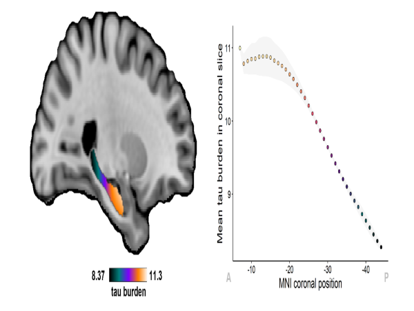
The researchers examined the distribution of deposits of tau and amyloid, two pathological proteins which are the hallmarks of Alzheimer's disease. For that purpose, they used valuable samples from Alzheimer’s patients collected with support of the Reina Sofia Foundation, in Madrid. These 3D analyses unveiled distinct distributions for these two pathologies along the hippocampus: tau pathology peaked at the anterior (front) end, while amyloid spread across the hippocampus in an “inverted-U” fashion. Amyloid pathology also displayed more complex gradients in other directions, with variable deposition patterns across different subfields of the hippocampus.
The authors concluded that Path2MR provides valuable insights into the distribution of Alzheimer’s pathology, opening new possibilities for understanding the intricate ways in which this disease affects the human brain. “Path2MR is a versatile tool which can map to 3D any observation made under the microscope at hospitals or research institutions. This means we can localize cellular or pathological alterations with much higher precision”. says Diana Ortega-Cruz, the first author of this study. In the field of Alzheimer’s, such 3D analyses can inform about the interaction between pathologies and their downstream effects, informing future treatment and diagnostic options.
Link to the complete article: https://alz-journals.onlinelibrary.wiley.com/doi/10.1002/alz.13695
Bryan Strange
Universidad Politécnica de Madrid
[email protected]
Visit us on social media:
Twitter
Other
![]()
